The Nervous Heart: Insights into Autonomic-Mediated Arrhythmias
Cardiac Imaging Data Analysis using BV Workbench
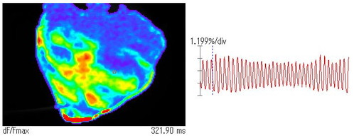
High Speed Imaging of Atrial and Ventricular Action Potential
- Animal
- Mouse
- Sample
- Isolated heart
- Dye
- Voltage Sensitive Dye
- Imaging System
- MiCAM03-N256
- Pixels
- 256×256
- Frame Rate
- 1,000fps (1.0msec/frame)
Ventricular Fibrillation in Pig Heart
- Animal
- Pig
- Sample
- Isolated Heart
- Dye
- Voltage Sensitive Dye
- Imaging System
- MiCAM03-N256
- Pixels
- 256×256
- Frame Rate
- 1,818fps (0.55msec/frame)
- Provided by
- Dr. Jack M. Rogers, The University of Alabama at Birmingham
Ventricular Fibrillation in Pig Heart
- Imaging System
- MiCAM02-HR
- Provided by
- Dr. Mihaela Pop, Sunnybrook Research Institute, Dept Medical Biophysics, University of Toronto
Simultaneous Calcium and Voltage Imaging in Human Ventricle

Simultaneous calcium and voltage imaging in human ventricle with MiCAM ULTIMA-L dual camera system. Figure shows (left to right): photo of transnural wedge human heart preparation, single pixel recordings of Vm and Ca, maps of action potential duration and calcium transient duration at 80% amplitude.
- Provided by
- Dr.Qing Lou and Dr.Igor Efimov, Washington University in St.Louis
Ventricular Fibrillation in Guinea Pig Heart (MiCAM ULTIMA-L)
- Traces of raw data [JPEG: 196KB]
- VF first derivative animation at 2,000 fps [MPEG4: 1.3MB]
Ventricular fibrillation (VF) in guinea pig heart. VF was induced by burst stimulation (35 ms interval, 100 pulses) and field of view is exactly 1×1 cm (1:1 magnification) from anterior region of heart. The first derivative of action potential was calculated to detect wave fronts as shown in this trace. The animation of action potential propagation clearly shows details of the direction and the curvature of the wave front. The heart was retrogradely perfused at 70 mmHg perfusion pressure and 20 ul of di-4 ANEPPS stock solution (1 mg / 1 mL DMSO).
- Provided by
- Dr.Bum-Rak Choi, Cardiovascular Research Center, Rhode Island Hospital and Brown Medical School
高速イメージングシステム MiCAM03-N256
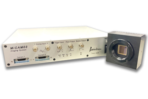
これまで世界中の脳神経/心臓研究分野で使用され、多くの実績を残したブレインビジョン製MiCAMシリーズの標準モデルである高速イメージングシステムMiCAM02の後継機種となります。
高速撮影・高S/N比、直感的な操作方法などの基本性能は継承しつつ、新開発CMOSカメラヘッドによる高画素化、高速データ転送による長時間記録などの新たな特長が加えられ、これまでには無いイメージングシステムへと進化を遂げました。